
Melanoma
- BONUS Official

- May 8, 2024
- 1 min read
Melanoma is the deadliest form of skin cancer and is the 5th most common cancer in the UK. Melanoma is not just a skin cancer, but can develop anywhere on the body, including the eyes, scalp, nails, feet and mouth. It can also spread widely to other parts of the body.

Definition"The abnormal and uncontrolled growth and division of melanocyte (melanin-producing cells)"
Epidemiology16,700 people are diagnosed with melanoma skin cancer in the UK each year.
It is the 5th most common cancer in the UK.
Risk Factors / CausesAge (younger for superficial spreading melanoma & older in other melanoma types)
Type 1 skin (caucasian, fair with freckles)
Ultraviolet light
Repeated sunburns
Intermittent sun exposure
Sun beds
Equatorial latitudes
Multiple moles (> 50-100 and > 5 mm)
Family history (e.g., CDKN2A mutations)
Giant congenital melanocytic naevus birthmark
Immunosuppression
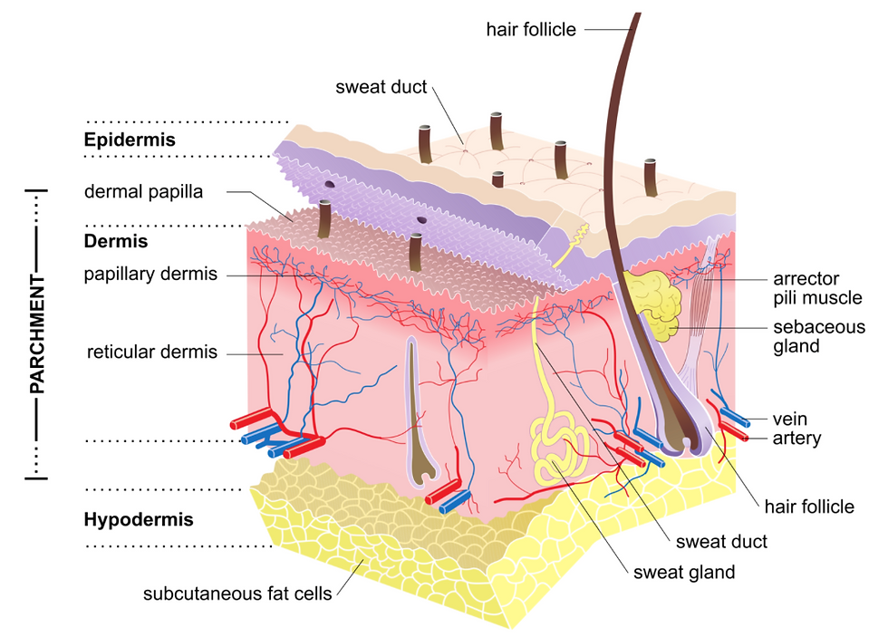
Anatomy / PathophysiologyThe skin has 3 broad layers: (1) epidermis, (2) dermis, and (3) hypodermis
Melanocytes are found within the deep part of the epidermis
These cells are responsible for melanin production and pigment formation
Individuals with darker skin tones do not have more melanocytes, their melanocytes are just more active
Melanocytes make greater amounts of melanin on sun exposure
The pigment helps to protect the body from ultraviolet light (UV radiation) from the sun and the DNA damage it can cause, leading to cancer development
There are several types of melanoma:
[1] Superficial spreading melanoma
Most common type of melanoma (70-80%)
Affects younger patients (30-50 years)
Present on arms, legs or torso
[2] Nodular melanoma
2nd most common type of melanoma (9-15%)
Most aggressive form of melanoma, worst prognosis as it metastasises early
Red/black lesions that bleed or weep

[3] Lentigo maligna melanoma
3rd most common type of melanoma
'In situ' melanoma confined to the epidermis (slowly growing coloured patches of skin)
Usually affects older caucasian individuals with chronic sun exposure
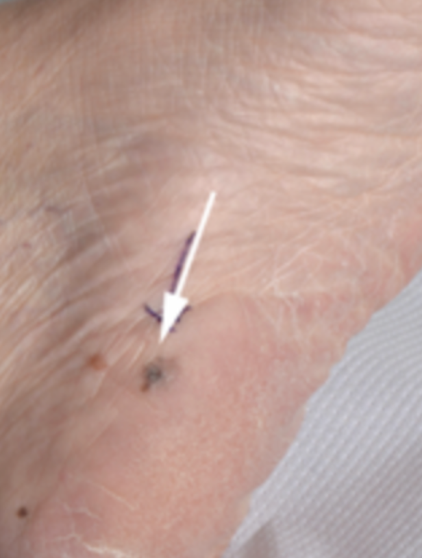
[4] Acral lentiginous melanoma
1-3% of melanoma cases
Affects the palms of the hands, soles of the feet or nail beds (longitudinal lines)
Most common type of melanoma in people with brown or black skin
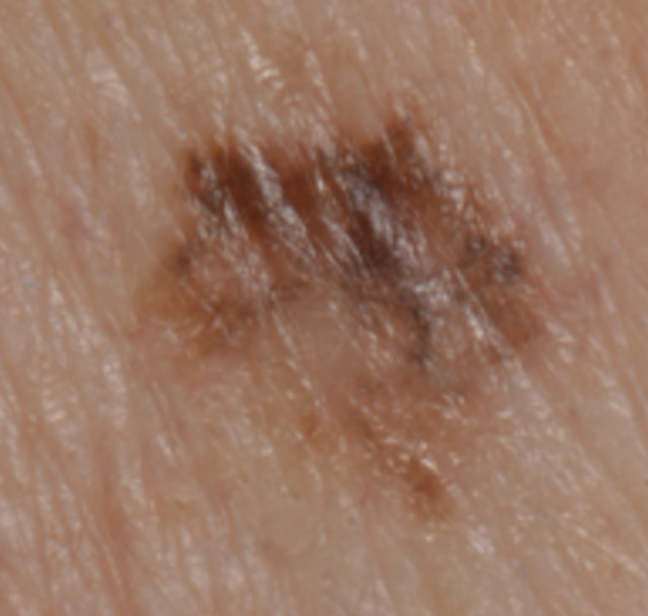
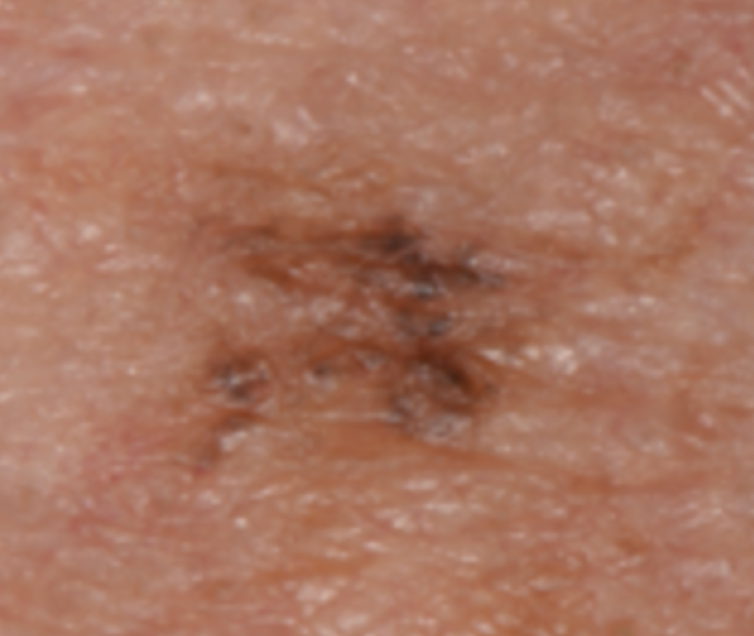
Signs and SymptomsSigns:
ABCDE:
A - Asymmetry to lesion
B - Border irregularity (poorly demarcated)
C - Colour variability and changing colour over time
D - Different to other melanocytic nevi/moles
E - Evolution (grow and change rapidly)
InvestigationsDermoscopy - magnifies lesion for clinicians to observe for features of melanoma
Excision biopsy - the lesion is cut out and sent for histological analysis to confirm diagnosis and grading
Lymph node biopsy - staging the melanoma to look for other cancerous lesions in lymph nodes
CT scan or MRI scan - staging the melanoma to look for other cancerous lesions in the body
BRAF V600 genetic testing - to help decide which treatment is best (targeted cancer therapy or immunotherapy)
Staging / GradingThe TNM staging system is a universal standard for classifying the extent of cancer. There are 3 components in this system:
Tumour (T): size and extent of the primary tumour
Node (N): regional lymph node involvement
Metastasis (M): presence of metastatic spread
As the TNM staging system is updated regularly, we recommend you stick to the version that your hospital uses. You can find the information about the TNM staging system version 8.0 here: https://www.cancerresearchuk.org/about-cancer/melanoma/stages-types/tnm-staging
ManagementSurgery
Radiotherapy
Chemotherapy
Immunotherapy
Targeted therapy
Intralesional therapy (injecting treatment directly into the melanoma e.g., talimogene laherparepvec or T-VEC)
Laser surgery (carbon dioxide laser)
Referral Criteria2-week-wait referral criteria:
A suspicious pigmented skin lesion with a score of ≥ 3 from the criteria below:
Major lesion features (scoring 2 points each):
Change in size
Irregular shape
Irregular colour
Minor lesion features (scoring 1 point each)
Largest diameter 7 mm or more
Inflammation
Oozing
Change in sensation
OR if dermoscopy suggests melanoma
OR people with a pigmented or non-pigmented skin lesion that suggests nodular melanoma
Resourceshttps://www.cancerresearchuk.org/about-cancer/melanoma/about
https://www.cancerresearchuk.org/about-cancer/melanoma/risks-causes
https://www.cancerresearchuk.org/about-cancer/melanoma/stages-types/types
https://www.cancerresearchuk.org/about-cancer/melanoma/stages-types/tnm-staging
https://www.cancerresearchuk.org/about-cancer/melanoma/treatment/treatment-decisions


Comments